Cell structure
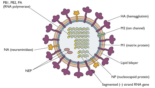
The influenza virion is a roughly spherical, enveloped virus. Glycoproteins, which look like spikes, are inserted into its lipid membrane. These proteins, HA (hemagglutinin) and NA (neuraminidase), determine the subtype of influenza virus and are important in the immune response against the virus. Antibodies against these spikes may protect against infection. (Racaniello)
Analysis of the sequences for receptor-binding and cleavage sites of hemagglutinin (HA), stalk region of neuraminidase (NA), non-structural protein 1 (NS1), polymerase basic protein 2 (PB2), and polymerase basic protein 1 (PB1) suggested that
Analysis of the sequences for receptor-binding and cleavage sites of hemagglutinin (HA), stalk region of neuraminidase (NA), non-structural protein 1 (NS1), polymerase basic protein 2 (PB2), and polymerase basic protein 1 (PB1) suggested that
- H1N1 a low virulent and low pathogenic virus
- Its replication is restricted to the cells of upper respiratory tract, so it does not lead to a systemic infection,
- It spreads among humans only
- Its replication could be inhibited by oseltamivir, zanamivir, interferon (IFN), and tumor necrosis factor alpha (TNF-alpha)." (Molecular Characteristics of the Human Pandemic Influenza A Virus (H1N1))
Genetics
H1N1 is a new hybrid strain of virus, which combines genetic materials from swine, human, and avian flu sources. The smallest protein in the influenza virus, 7 PB1-F2 coding sequence, is the known molecular marker of pathogenicity as it is exclusively absent in human influenza viruses. The second marker is the degree of identity between the viral hemagglutinin molecules of new strain and other human flu viruses that can be assessed by sequences alone. Low identity of hemagglutinin structures indicates that degree of “herd” immunity resulting from exposure to similar viruses does not blunt the transmission from one human to another. Polybasic cleavage site, which is a protease site in the viral hemagglutinin, plays a role as the third molecular marker in the pathogenicity of avian influenza viruses. These host proteases enables virus fusion with a host cell by activating the hemagglutinin molecule. (Cazzola)
Pathogenisis
There is no evidence of true long-term carrier state. After inhalation, H1N1 virus is deposited on the surface of the lower respiratory tract.
Uncomplicated infections may alter cranial ventral lung lobes while bronchial and mediastinal lymph nodes become enlarged.
Notice pathological changes:
Uncomplicated infections may alter cranial ventral lung lobes while bronchial and mediastinal lymph nodes become enlarged.
Notice pathological changes:
- Sharp line of demarcation between normal and affected lung tissue can be identified with the affected tissue being purple and firm.
- Interlobular edema can be found in few cases.
- Airways get filled up with blood-tinged fibrinous exudates with peribronchial and perivascular cellular infiltration.
- Fibrinous pleuritis is seen in severe cases.
- Microscopically lesions show airways filled with exudate, with extensive alveolar atelectasis, interstitial pneumonia and emphysema.
- Research revealed that widespread interstitial pneumonia prevails up to 21 days after infection and causes hemorrhagic lymph nodes (Cazzola)
